Veins from zebrafish make arteries get a move on
Max Planck scientists in Münster develop a procedure that allows unique insight into vessel growth
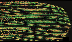
The development of early embryonic blood vessels follows a strictly organized pattern: the anatomy of the first large arteries and veins is almost identical between various individuals. Later in development, but also in wound healing, new arteries and veins are mostly formed in an intermediate step; a so-called vascular plexus is established - a disorganized network of blood vessels. It was largely unknown how exactly these arteries and veins are formed from this plexus, because tissue of vertebrates is mostly non-transparent. This makes it hard to film cell movements. Dr. Arndt Siekmann and colleagues of the Max Planck Institute for Molecular Biomedicine have now developed a procedure by which they could film blood vessel growth in adult zebrafish for over one day. The scientists observed that new blood vessels predominantly sprout from veins. They further discovered that some cells in this network built an arch and then migrated backwards within the expanding tissue. Interestingly, exactly these cells later constituted the arteries (Nature Communications, 15. December 2014).