Embryo Self-Correction
Rocha Lab
Exploring how embryos overcome developmental defects to form functional organs
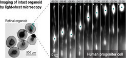
In nature, embryos frequently encounter genetic and environmental stressors (e.g., mutations, hypoxia, and viral infections) that cause developmental defects. Given these significant challenges, it is truly remarkable that embryos typically develop healthy organs. So far, strong emphasis has been placed on the identification of the molecular basis of developmental robustness; however, how developing tissues restore cellular order after severe disturbances remains an important knowledge gap. To shed light on how cells adapt and work together to provide self-correction capabilities, we challenge developing tissues with diverse stressors, including physical stressors and disease variants, that disrupt cell behaviour. Using advanced live imaging techniques, we closely interrogate how developing tissues “break” and “fix” themselves, focusing on regrowth and repair of tissue organization. We aim to dissect mechanisms of self-correction from the molecular to the tissue scale by combining quantitative imaging with large-scale molecular profiling and biophysical measurements. We hope to deepen our understanding of how embryos build healthy organs even in conditions that could cause disease.
Methodological and technical expertise

- Human organoid models
- Zebrafish genetics (e.g., CRISPR)
- Advanced microscopy and deep-learning image restoration tools (e.g., light-sheet microscopy, CARE)
- Quantitative image analysis (e.g., Python, Fiji)
